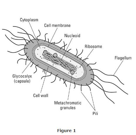
Until light microscopes with better lenses and electron microscopes with higher magnifying capabilities were developed, microbiologists knew little about the structure and considerably more about the chemistry of the organisms they studied. A typical bacterial cell is illustrated in Figure with the major features named. The cell is most of the features found in eukaryote cells.

When growth conditions become unfavorable—when nutrients become scarce, for example, or the environment dries—many bacteria produce endospores adding thick walls around the circular DNA together with a bit of cytoplasm. The spores resist high temperatures, desiccation, chemical disinfectants, ultraviolet radiation, X‐rays, boiling for several hours and are the reason bacteria sometimes survive even in “sterile” hospital environments.
Bacteria are basically unicellular with simple shapes: short rods or bacilli (singular: bacillus), spheres or cocci, or spiral, elongated cells, spirilla. The single cells often are linked together into ribbon‐like filaments, or bead‐like chains of cells; some taxa form flat, sheet‐like colonies, others produce stalked, branching ones.
All except the mycoplasmas have a cell wall composed of disaccharides and peptides (amino acids) together with a unique compound not found in eukaryotes: peptidoglycan. The latter substance is present in the Domain Bacteria and absent in the Domain Archaea, making it a good diagnostic feature. The gram stain, a dye that reacts with peptidoglycan and proteins of the cell walls, effectively divides the Domain Bacteria individuals into two major groups, gram‐positive and gram‐negative members. (In the days of light microscopes when not much beyond general rod‐coccus‐spirilla shape was discernable, bacteriologists struggled to find good diagnostic features; the chemistry of the walls proved to be one.)
|
|
|
|
|
|
|
|
|
|
|
|
|
|
|
|
|
|
|
|
|
|
|
|
|
|
|
|
|
|
|
|
|
|
|
|
|
|